|
СЛЁЗНЫЙ АППАРАТ [ nasolacrimal apparatus ] Слёзный аппарат - это части органа зрения, относящиеся к вспомогательным структурам глаза.
К слёзному аппарату относят совокупность следующих структур: слёзная железа, выводные канальцы слёзной железы, конъюнктивальный мешок, в который открываются выводные канальцы слёзной железы, слёзоотводящие пути.
ОРГАН ЗРЕНИЯ: ОГЛАВЛЕНИЕ = organ of vision: content
- ВСПОМОГАТЕЛЬНЫЙ АППАРАТ ГЛАЗА – accessory visual apparatus
- ВЕКИ – eyelids, palpebrae
- КОНЪЮНКТИВА ВЕК И ГЛАЗНОГО ЯБЛОКА – conjunctiva of eyelids and eyeball
- ГЛАЗНИЦА – eye's orbit
- КОСТНАЯ ОСНОВА ГЛАЗНИЦЫ – bones of the eye's orbit
- МЯГКИЕ СОЕДИНИТЕЛЬНОТКАННЫЕ СТРУКТУРЫ ГЛАЗНИЦЫ – soft connective tissue within the eye orbit
- ВНЕШНИЕ МЫШЦЫ ГЛАЗНОГО ЯБЛОКА – extrinsic (extra-ocular) muscles
- СЛЁЗНЫЙ АППАРАТ – nasolacrimal apparatus
- КРОВЕНОСНЫЕ СОСУДЫ ГЛАЗНИЦЫ – blood vessels within the eye orbit
- ЛИМФАТИЧЕСКИЕ СТРУКТУРЫ ГЛАЗНИЦЫ – limphatic structures within the eye orbit
- НЕРВЫ ГЛАЗНИЦЫ – nervs within the eye orbit
- ГЛАЗ – eye
- ГЛАЗНОЕ ЯБЛОКО – eyeball
ОРГАН ЗРЕНИЯ: ОГЛАВЛЕНИЕ
ОРГАН ЗРЕНИЯ: ТАБЛИЦЫ
ОРГАН ЗРЕНИЯ: ИЛЛЮСТРАЦИИ
ОРГАН ЗРЕНИЯ: ЛИТЕРАТУРА
СЛЁЗНЫЙ АППАРАТ.
К слёзному аппарату относят совокупность следующих структур: слёзная железа, выводные канальцы слёзной железы, конъюнктивальный мешок, в который открываются выводные канальцы слёзной железы, слёзоотводящие пути.
Cлёзная железа - это сложная дольчатая альвеолярно-трубчатая железа. Она расположена в одноименной ямке латерального угла верхней стенки глазницы. Cлёзная железа разделяется на две части, проходящим через неё сухожилием мышцы, поднимающей верхнее веко. Большая часть железы - её верхняя глазничная часть. Меньшая часть железы - её нижняя вековая (пальпебральная) часть лежит возле верхнего свода конъюнктивы. Под сводом конъюнктивы иногда находятся небольших размеров добавочные слёзные железы.
Выводные канальцы слёзной железы (приблизительно ~15 канальцев) открываются в конъюнктивальный мешок. Конъюнктивальный мешок расположен в латеральной части верхнего свода конъюнктивы. Слёзная жидкость (слеза), поступающая из слёзной железы по канальцам, омывает переднюю поверхность глазного яблока. Избыток слёзной жидкости оттекает по слёзному ручью (капиллярная щель возле краёв век) в область медиального угла глаза, в слёзное озеро. В этом месте берут начало короткие (длиной ~1 см) и узкие (диаметром ~0,5 мм) верхний и нижний слёзные канальцы. Эти изогнутые канальцы, раздельно или соединившись друг с другом, открываются в слёзный мешок. Cлёзный мешок расположен в одноименной ямке в нижнемедиальном углу глазницы. Книзу слёзный мешок продолжается довольно широким (до ~4 мм) носослёзным протоком. Носослёзный проток заканчивается в носовой полости, в передней части нижнего носового хода. Передняя стенка слёзного мешка сращена со слёзной частью круговой мышцы глаза. При сокращении слёзной части круговой мышцы глаза слёзный мешок расширяется. Это расширение слёзного мешка приводит к всасыванию слёзной жидкости через слёзные канальцы в слёзный мешок.
Franz H. Grus, Stephanie C. Joachim and Norbert Pfeiffer Proteomics in ocular fluids Proteomics Clin. Appl. 2007, 1, 876–888
2_184/Proteomics in ocular fluids2007.pdf
Whikehart D.R., Ed. Biochemistry of the Eye, 2nd ed., Elsevier, 2003, 331 p.
2_184/Biochemistry of the Eye2ed2003.pdf
|
Схема. Веки и глазное ябоко.
Модификация: Gray H., (1821–1865), Standring S., Ed. Gray's Anatomy: The Anatomical Basis of Clinical Practice. 39th ed., Churchill Livingstone, 2008, 1600 p.
см.: Анатомия человека: Литература. Иллюстрации.
Примечание:
Веки расположены кнаружи от передней поверхности глазного яблока. Они частично или полностью прикрывают поверхность глазного яблока сверху и снизу. Неприкрытое веками пространство между их свободными краями называется глазной щелью. При смыкании век они могут замкнуть глазную щель и полностью прикрыть переднюю поверхность глазного яблока. Ширина и форма глазной щели может иметь индивидуальные и межиндивидуальные изменения. |
 |
|
|
Примечание:
|
 |
|
|
Схема. Секреторная единица слёзной железы.
Модификация: Gray H., (1821–1865), Standring S., Ed. Gray's Anatomy: The Anatomical Basis of Clinical Practice. 39th ed., Churchill Livingstone, 2008, 1600 p.
см.: Анатомия человека: Литература. Иллюстрации |
 |
|
|
Схема. Верхнее веко и передний сегмент глаза. Сагиттальное сечение.
Модификация: Gray H., (1821–1865), Standring S., Ed. Gray's Anatomy: The Anatomical Basis of Clinical Practice. 39th ed., Churchill Livingstone, 2008, 1600 p.
см.: Анатомия человека: Литература. Иллюстрации |
 |
|
Примечание:
|
• Генле Фридрих (Henle Friedrich Gustav Jacob, 1809–1885) — германский анатом и патолог.
• Генле Фридрих (Henle Friedrich Gustav Jacob, 1809–1885) — анатом, патолог, Германия.
• Краузе Карл (Krause Karl Friedrich Theodor, 1797–1868) — анатом, врач, Германия.
• Мейбом Генрих (Meibom Heinrich, 1638-1700) - анатом, Германия.
• Молль Якоб (Moll Jacob Antonius, 1832-1914) - анатом, офтальмолог, Голландия.
• Мюллер Генрих (Müller Heinrich, 1820–1864) — анатом, Германия.
• Риолан Жан (Riolan Jean, 1577-1657) - анатом, врач, Франция.
• Цейс Эдуард (Zeis Eduard, 1807-1868) - врач, Германия. |
|
|
Схема. Слои плёнки предкорнеальной слёзной жидкости.
Модификация: Levin L.A., Nilsson Siv F.E., Ver Hoeve J., Wu S., Kaufman P.L., Alm A. Adler's Physiology of the Eye. Elsevier, 2011, 820 p. см.: Физиология человека: Литература. Иллюстрации. |
 |
|
Примечание:
|
The lipid layer is secreted by the meibomian glands, modified sebaceous glands, that line the upper and lower eyelids in a single row.[52,53] A single meibomian gland consists of multiple acini that secrete into ducts that converge to form a common duct that exits onto the eyelid near the mucocutaneous junction (see Fig. 15.1). The acini are surrounded by nerves and blood vessels. The cells in the acini are arranged in a specific order that reflects their function such that the outer cell layer consists of undifferentiated, flattened basal cells. As these cells mature they move inward to the center of the acinus and concomitant with this inward migration is continuing synthesis of lipids. Meibomian gland lipids are stored in vesicles. As the cells move closer to the center they contain increasingly more lipid secretory vesicles reflecting continued lipid synthesis. Upon the appropriate stimulus (unknown), the cells in the acinus center burst, releasing the entire cell contents in their secretory granule lipids and the other components of the cell into the duct system. This releases the entire cell contents and is known as holocrine secretion. The secreted cells are replaced by proliferation of the basal cells.[54] Thus the secretory product contains a complex mixture of lipids and proteins and is termed meibum. The synthesized lipids are the major component of meibum, with the minor lipid classes probably reflecting the cellular membrane lipids. Meibomian fluid is a mixture of non-polar lipids (wax esters, cholesterol, and cholesterol esters) and polar lipids (mainly phospholipids). Meibum is liquid at lid temperature.
The secreted lipid is stored in the duct system that terminates in orifices, with a muscular cuff, that open onto the lid.[53] Meibum is released on to the ocular surface in small amounts with each blink forming a casual reservoir with about 30 times more lipid than needed for each blink. With the up-phase of each blink the upper lid draws oil from the lid reservoir and spreads it over the anterior tear film surface. With the down phase the lipid film is returned to the marginal reservoir as the lid closes.[53] The lipids mix only slightly with the lipid reservoir as it folds up as an intact sheet providing a gradual turnover of the lipids in the tear film.
Butovich et al[55] have proposed that proteins and mucins contribute to the lipid layer so that the proteins, which contain hydrophobic, hydrophilic, and charged portions, can unfold and form a variety of shapes depending upon the local milieu (Fig. 15.15). The proteins could extend across the lipid layer suggesting a complex mixture of islands of proteins, mucins, and lipids, similar to that of lung surfactant. This model would be a non-collapsible, viscoelastic gel and allow for the lowest free energy states of the proteins in contact with lipids. Changes in the osmolarity of tears would affect the ability of the proteins to unfold and interact with the lipids. This model also accounts for the lack of change of interference patterns after each blink. |
|
Таблица. Сравнение концентраций некоторых компонентов крови и водянистого слоя слёзной жидкости = Comparison of some components of blood with aqueous layer tears.
Модификация: Whikehart D.R., Ed. Biochemistry of the Eye, 2nd ed., Elsevier, 2003, 331 p., см.: Физиология человека: Литература. Иллюстрации. |
№ |
Component |
Blood Concentration |
Tear Concentration |
| 1 |
Ascorbate |
1,3 mg / 100 ml |
0,4 mg / 100 ml |
| 2 |
Bicarbonate |
27 mmol / liter |
23 mmol / liter |
| 3 |
Calcium |
4,8 mg / 100 ml |
1 mg / 100 ml |
| 4 |
Glucose |
90 mg / 100 ml |
6 mg / 100 ml |
| 5 |
Potassium |
4,3 mmol / liter |
30 mmol / liter |
| 6 |
Protein |
7 g / 100 ml |
0,7 g / 100 ml |
| 7 |
Sodium |
150 mmol / liter |
138 mmol / liter |
|
Таблица. Липиды слёзной жидкости.
Модификация: Whikehart D.R., Ed. Biochemistry of the Eye, 2nd ed., Elsevier, 2003, 331 p., см.: Физиология человека: Литература. Иллюстрации. |
№ |
Lipid Component |
Composition (%) |
| 1 |
Cholesteryl esters |
29,5 |
| 2 |
Wax esters |
35 |
| 3 |
Triacylglycerols |
4 |
| 4 |
Cholesterol |
1,8 |
| 5 |
Fatty acids |
2,2 |
| 4 |
Unidentified1 |
27,5 |
|
====
Слезная жидкость - водянистая жидкость, которая, как показывает табл. 5.1 , не сильно отличается по составу от плазмы , за исключением намного более высокой концентрации К+ и Cl- и отсутствия большей части органики. В слезной жидкости присутствует множество ферментов, наиболее важный из них - лизоцим , который атакует бактерии, растворяя их клеточные стенки. Слезная жидкость двигается по поверхности роговицы в результате мигательных движений и дренируется в носовом углу глаза системой, в которую входят слезное озеро, канальцы, слезный мешок и носо-слезный проток, ведущий в носовую полость ( рис. 15.4 ). Кроме того, у человека выделение слезной жидкости служит для выражения эмоций.
Предроговичная жидкость, или слёзная жидкость - это прозрачная водянистая жидкость со слабощелочной реакцией (рН ~7,0 - 7,4). При нормальном состоянии многокомпонентная слёзная жидкость вырабатывается в небольших количествах (~0,5-1,0 мл в сутки) непрерывно. При закрытых веках слёзная жидкость заполняет щелевое пространство между веками и глазным яблоком. При открытых веках слёзная жидкость в течение ограниченного времени (~15 с) в виде плёнки покрывает наружную поверхность глазного яблока, его роговицу.
Слёзная жидкость состоит из трёх компонентов. Наибольшим по объему компонентом является водянистый раствор белков, солей, растворимых муцинов и некоторых других веществ. Этот компонент вырабатывается слёзными железами и называется слезой. Концентрации растворённых веществ водянистого компонента слёзной жидкости в сравнении с плазмой крови представлены в таблице ниже. Слеза содержит ряд ферментов. Наиболее изученным ферментом является лизоцим. Вторым компонентом слёзной жидкости являются липиды. Липидный секрет вырабатывается главным образом мейбомиевыми желёзами, желёзами Цейса и желёзами Молля. Состав липидов липидного слоя слёзной жидкости представлен в таблице ниже. Третьим компонентом слёзной жидкости являются муцины. Муциновый секрет вырабатывается главным образом бокаловидными клетками конъюнктивы, клетками крипт Генле конъюнктивы, железами Манца конъюнктивы. Описанные компоненты образуют сложную частично регулярную структуру - плёнку слёзной жидкости.
Плёнка слёзной жидкости состоит из трёх слоёв. Наружный слой (со стороны среды, атмосферы) представляет собой слой молекул липидов, липидный слой слёзной жидкости (толщина ~3-100 нм).
Липидный слой замедляет испарение воды из слёзной жидкости. Толщина липидного слоя при открытом глазе составляет в среднем ~10 нм (~13-170 нм). Толщина липидного слоя зависит от ряда факторов. Среди них:
• интенсивность секреции мейбомиевых желёз;
• состав липидных секретов желёз;
• частота и эффективность моргания век, распределяющих слёзную жидкость по поверхностям глазного яблока;
• площадь поверхности глазной щели между свободными краями век;
• влажность среды человека.
Внутренний слой (прилежащий к эпителию роговицы) - это слой муцина, муциновый слой слёзной жидкости (толщина ~0,002 - 0,005 мкм). Средний слой, водянистый слой слёзной жидкости (толщина ~3-40 мкм), основной по объёму, представляет собой жидкий раствор, растворителем в котором является вода (~98-99 %). В воде равномерно распределены различные вещества в виде частиц, молекул или ионов.
В результате мигательных движений век слёзная жидкость распределяется по поверхности роговицы. Излишки слёзной жидкости стекают в капиллярную щель между задним ребром нижнего века и глазным яблоком. В виде слёзного ручейка слёзная жидкость попадает в носовой угол глазницы. Из носового угла глазницы слёзная жидкость поступает в дренажную носослёзную систему (последовательно: слёзное озеро, слёзные канальцы, слёзный мешок и носослезный проток, открывающийся в носовой полости). Продвижению слезы в сторону дренажной носослёзной системы способствуют мигательные движения век.
Слёзная жидкость участвует в осуществлении ряда важных функций:
• механическая (удаление пылевых частиц, вредных веществ) и антибактерицидная защита;
• оптимизация оптической среды глаза (сглаживает микроскопические неровности поверхности роговицы, обеспечивает её влажность, гладкость и зеркальность);
• участие в дыхании и питании роговицы.
. Кроме того, у человека выделение слезной жидкости служит для выражения эмоций.
Beuerman R.W. The Lacrimal Functional Unit = Лакримальная функциональная единица. p. 11-39, In: Pflugfelder S., Ed. Dry Eye and Ocular Surface Disorders = Нарушения стабильности предроговичной слёзной плёнки. Сухой глаз. Marcel Dekker, 2004, 439 p.
Louisiana State University Eye Center, New Orleans, Loьisiana, U.S.A.,
and Singapore Eye Research Institute, Singapore
Austin Mircheff
University of Southern California Keck School of Medicine,
Los Angeles, California, U.S.A.
Stephen C. Pflugfelder
Baylor College of Medicine, Houston, Texas, U.S.A.
Michael E. Stern
Allergan, Inc., Irvine, California, U.S.A.
!!!!! Pflugfelder S., Ed. Dry Eye and Ocular Surface Disorders = Нарушения стабильности предроговичной слёзной плёнки. Сухой глаз. Marcel Dekker, 2004, 439 p. Обзоры, физиология
Stephen (Editor), Roger Bauerman (Editor), Michael E. Stern (Editor)
6_117/Ocular Surface2004.pdf
NORMAL ANATOMY AND PHYSIOLOGY
Tears function in the following ways to maintain ocular health:
Cover minor epithelial surface irregularities, making the cornea a smooth optical surface
Moisten and protect delicate surfaces of the corneal and conjunctival epithelium
Nourish the avascular corneal tissues
Inhibit microbial growth by mechanical flushing and antimicrobial action
The tear film consists of three separate layers, each serving a unique function: (Refer to Figure 1)
The outermost lipid layer, derived from meibomian glands in the eyelid margins, contains cholesterol, fatty acids and triglycerides. It retards evaporation of the aqueous tear layer.
The middle aqueous layer is produced by lacrimal and accessory glands. It constitutes approximately 98% of the tear film and contains water soluble substances such as electrolytes, proteins, nutrients and immunoglobulins.
The innermost mucin layer is composed of various glycoproteins produced by goblet cells, which are specialized epithelial cells on the corneal and conjunctival surfaces of the eye. Some glycoproteins attach to microvilli on the corneal and conjunctival epithelial cells, providing a cushiony surface that lubricates the surface of the eye. Other glycoproteins secure the aqueous tear layer, allowing it to spread over the mucin layer.
#2
Parker J., Parker P., Eds. Dry Eyes - A Medical Dictionary, Bibliography, and Annotated Research Guide to Internet References ICON Health Publications, 2004, 104 p.
6_117/Dry_Eyes_Med_Diс2004.pdf
The composition of lipids in the tears is rather complex. A wide
variety of fatty acids, their alcohol derivatives, and cholesterol make up
the waxes and cholesteryl esters that are the majority of lipids found
there. Other lipid classes are also present as described by Nicolaides
(1986) and as shown in Table 5–4. Slightly more than 25% of the lipid
types have still not been completely characterized. They include 8.4%
double esters or diesters (Figure 5–18) in which hydroxy fatty acids are
esterified to two other fatty acid(s) or an alcohol or even a cholesterol
molecule. Four percent of the lipids represent uncombined, precursor
fatty acids and cholesterol molecules. The varieties of fatty acids are
listed in Table 5–5. Nicolaides has estimated that with some 69 different
fatty acids, 40 fatty acid alcohols and 11 hydroxy fatty acids in
Meibomian gland secretions, about 30,000 ester species are possible!
The cooperative physical-chemical properties of these esters provide for
the ability of the lipids: (1) to flow from their ducts to the eyelid edges;
(2) to form a film over the aqueous layer and maintain contact with it;
(3) to adhere to the eyelid skin and act as a barrier to the aqueous layer;
and (4) to form a water-tight seal when the lids are closed.
Pathological conditions can alter this exquisite ester mixture and
bring about tear film abnormalities. In Meibomian gland dysfunction,
Lysozyme
Lysozyme is an enzyme of the precorneal tear film that is instrumental in
destroying certain kinds of bacteria (namely, those which are positively
stained with a crystal violet-iodine complex for peptidoglycans known as
Gram stain). These Gram-positive bacteria possess an outer coat of a
peptideglycan (sugar) polymer (or peptidoglycan), which, in Gramnegative
bacteria, is only transiently stained since those bacteria are
covered up by a second, outer lipid membrane. Lysozyme is able to
hydrolyze or break up the glycan (sugar polymer) components of the
peptidoglycan of Gram-positive bacteria as shown in Figure 3.13. The
enzyme was initially described in 1922 by Alexander Fleming, a British
bacteriologist (Stryer, 1988). He first found it in nasal mucous, but later
discovered that tears are a rich source of the enzyme. The concentration
has been estimated at 1.3 mg per ml of tears, which are unstimulated by
external or internal sources such as onion odor or emotional stress (Sen
and Sarin, 1980). Specifically, the enzyme breaks the ѓА1ЃЁ4 glycosidic
bond of the oxygen bridge between the repeating glycan units of
N-acetylmuramic acid (NAM) and N-acetylglucosamine (NAG) as
indicated in Figure 3.14. Lysozyme itself is a globular protein with a
molecular weight of about 14,000. A portion of the bacterial peptidoglycan
is able to fit in a groove on the outer face of the enzyme that contains
the active site (Figure 3–15). This is an enzyme in which the
detailed mechanism for hydrolysis is known and can be described in
three stages. Figure 3–16 shows the mechanism in which the disaccharide
unit is hydrolyzed. The active site contains two amino acid components
(Glu and Asp) whose carboxylate groups participate in the
hydrolysis. Initially, a proton (H+ from the Glu) breaks the bond by
binding to the oxygen between the two sugar rings leaving an unbound,
positively charged carbonium ion (carbon #4) in the right hand sugar
ring. This carbonium ion is temporarily stabilized by the negative charge
on Asp located above it (see Figure 3–16 A and B). Then, a nearby water
molecule ionizes and donates its proton to the negatively charged Glu
while the hydroxy group (–OH) binds to the carbonium ion and the reaction
is complete (see Figure 3–16 B and C). At completion, the original
forms of the enzyme are regenerated and the hydrolyzed (split) chains of
the peptidoglycan leave the active site of the enzyme.
Once the peptidoglycan cover is split open (hydrolyzed) by
lysozyme, the bacterium is no longer able to contain its high, internal
osmotic pressure with its plasma membrane alone and it bursts open.
Other protein components of tear film have also been implicated in bacteriocidal
action (Selsted, Martinez, 1982), but none of them are as
efficient as lysozyme.
In addition to its bacteriocidal activity, lysozyme also serves as an
important analytical indicator of tear dysfunction. Measurement of lysosomal
activity reflects the productivity of the main and accessory
lacrimal glands (Gillette, Greiner, Allansmith, 1981) as well as the status
of aqueous deficiency of the tear film (Van Bijsterveld, 1974; Van
Bijsterveld, Westers, 1980). In the application of a Micrococcus agar diffusion
assay (also known as a Schirmer Lysoplate Assay), for example,
tear film is collected on filter paper discs and placed on a dish with agar
containing 5 Ч 107 organisms of Micrococcus lysodeiticus. The lysozyme
in the tear sample is allowed to hydrolyze the peptidoglycans and destroy
the bacteria for 24 hours at 37°C. Then the diameter of the clear area of
agar (i.e., destroyed bacteria) surrounding the tear sample is measured
(Figure 3–17). This cleared diameter may be converted to units of
enzyme activity or simply interpreted either as normal or indicative of
tear dysfunction. More recently, Klaeger and coworkers have developed
a more reliable and efficient assay in which enzyme activity is measured
from collected tears with the substrate: p-nitrophenyl penta-N-acetyl
в-chitopentaoside. This substrate releases the colored product:
p-nitrophenol upon enzyme hydrolysis and tear sample lysozyme activity
can be analyzed within one hour.
СЛЕЗНЫЙ АППАРАТ (APPARATUS LACRIMALIS)
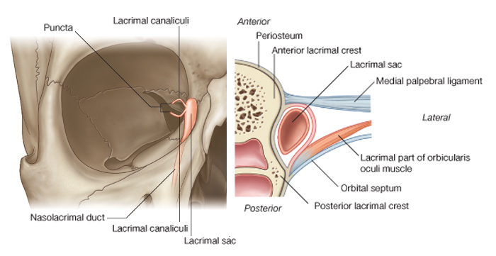
Собственно слезоотводящие пути состоят из слезных канальцев, слезного мешка и носослезного протока (рис. 18).
Слезные канальцы (canaliculi lacrimalis) начинаются слезными точками (punetum lacrimale), которые находятся на вершине слезных сосочков обоих век и погружены в слезное озеро. Диаметр точек при открытых веках 0,25-0,5 мм. Они ведут в вертикальную часть канальцев (длина 1,5-2,0 мм). Затем ход их меняется почти на горизонтальный. Далее они, постепенно сближаясь, открываются позади lig. palpebrale mediale в слезный мешок, каждый в отдельности или слившись предварительно в общее устье. Длина этой части канальцев 7—9 мм, диаметр 0,6 мм.
Стенки канальцев покрыты многослойным плоским эпителием, под которым находится слой эластических мышечных волокон.
Слезный мешок (saccus lacrimalis) расположен в костной, вытянутой по вертикали, ямке между передним и задним коленами lig. palpebrale
mediale и охвачен мышечной петлей (m. Horneri). Купол его выступает над этой связкой и находится пресептально, т. е. вне полости глазницы (см. рис. 9). Изнутри мешок покрыт многослойным плоским эпителием, под которым находится слой аденоидной, а затем плотной волокнистой тканей.
В нижнем отделе упомянутой ямки слезный мешок открывается в носослезный проток (ductus nasolaerimalis), который проходит сначала в костном канале (длина 9 мм). В нижнем же отделе он имеет костную стенку только с латеральной стороны, в остальных отделах граничит со слизистой носа и окружен богатым венозным сплетением. Открывается под нижней носовой раковиной в 3-3,5 см от наружного отверстия носа. Общая длина его 15 мм, диаметр 2-3 мм. У новорожденных выходное отверстие канала нередко закрыто слизистой пробкой или тонкой пленкой, благодаря чему создаются условия для развития гнойного или се-розно-гнойного дакриоцистита. Стенка канала имеет такое же строение, как и стенка слезного мешка. У выходного его отверстия слизистая оболочка образует складку (plica lacrimalis), которую Hasner описал как запирающий клапан.
В целом можно принять, что слезоотводящий путь состоит из небольших мягких трубок различной длины, формы и переменного диаметра, которые стыкуются под определенными углами. Они соединяют конъюнктивальную полость с носовой, куда и происходит постоянный отток слезы. Последний обеспечивается за счет мигательных движений век, сифонного эффекта с капиллярным притяжением жидкости, заполняющей слезные пути, перистальтического изменения диаметров канальцев, присасывающей способности слезного мешка (вследствие чередования в нем положительного и отрицательного давления при мигании) и отрицательного давления, создающегося в полости носа при аспираци-онном движении воздуха.
Продукция собственно слезы осуществляется слезной железой (glandula lacrimalis) и мелкими добавочными железками Краузе и Воль-фринга. Однако именно они обеспечивают суточную потребность глаза в увлажняющей его жидкости. Главная же слезная железа активно функционирует лишь в условиях эмоциональных всплесков (положительных и отрицательных), а также в ответ на раздражение чувствительных нервных окончаний в слизистой оболочке глаза или носа (рефлекторное слезоотделение).
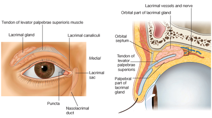
Слезная железа лежит под верхненаружным краем глазницы в fossa glandulae lacrimalis лобной кости (рис. 18). Сухожилие поднимателя верхнего века делит ее на большую глазничную и меньшую пальпебраль-ную части. Выводные протоки глазничной доли железы (в количестве
3-5) проходят между дольками пальпебральной железы, принимая попутно ряд ее многочисленных мелких протоков, и открываются в своде конъюнктивы в нескольких миллиметрах от верхнего края хряща. Кроме того, пальпебральная железа имеет и самостоятельные протоки, которых насчитывается от 3 до 9. Поскольку она лежит сразу же под верхним сводом конъюнктивы, то при вывороте верхнего века ее дольчатые контуры обычно хорошо видны.
Слезная железа иннер-вируется секреторными волокнами п. facialis, которые, проделав сложный путь, достигают ее в составе п. lacrimalis, являющегося ветвью п. ophthalmicus. У детей она начинает функционировать к концу второго месяца жизни. Поэтому до истечения этого срока при плаче их глаза остаются сухими.
|
Схема. Слёзная железа и мышца, поднимающая верхнее веко = Lacrimal gland and levator palpebrae superioris.
Модификация: Gray H., (1821–1865), Drake R., Vogl W., Mitchell A., Eds. Gray's Anatomy for Students. Churchill Livingstone, 2007, 1150 p.
см.: Анатомия человека: Литература. Иллюстрации |
 |
|
Примечание:
|
The lacrimal apparatus is involved in the production, movement, and drainage of fluid from the surface of the eyeball. It is made up of the lacrimal gland and its ducts, the lacrimal canaliculi, the lacrimal sac, and the nasolacrimal duct.
The lacrimal gland is anterior in the superolateral region of the orbit, and is is divided into two parts by the levator palpebrae superioris;
• the larger orbital part is in a depression, the lacrimal fossa, in the frontal bone;
• the smaller palpebral part is inferior to levator palpebrae superioris in the superolateral part of the eyelid.
Numerous ducts empty the glandular secretions into the lateral part of the superior fornix of the conjunctiva.
Fluid is continually being secreted by the lacrimal gland and moved across the surface of the eyeball as the eyelids blink.
Innervation
The innervation of the lacrimal gland involves three different components.
Sensory innervation
Sensory neurons from the lacrimal gland return to the CNS through the lacrimal branch of the ophthalmic nerve [V1].
Secretomotor (parasympathetic) innervation
Secretomotor fibers from the parasympathetic part of the autonomic division of the PNS stimulate fluid secretion from the lacrimal gland. These preganglionic parasympathetic neurons leave the CNS in the facial nerve [VII], enter the greater petrosal nerve (a branch of the facial nerve [VII]), and continue with this nerve until it becomes the nerve of the pterygoid canal).
The nerve of the pterygoid canal eventually enters the pterygopalatine ganglion where the preganglionic parasympathetic neurons synapse on postganglionic parasympathetic neurons. The postganglionic neurons join the maxillary nerve [V2] and continue with it until the zygomatic nerve branches from it, and travel with the zygomatic nerve until it gives off the zygomaticotemporal nerve, which eventually distributes postganglionic parasympathetic fibers in a small branch that joins the lacrimal nerve. The lacrimal nerve passes to the lacrimal gland.
Sympathetic innervation
Sympathetic innervation of the lacrimal gland follows a similar path as parasympathetic innervation. Postganglionic sympathetic fibers originating in the superior cervical ganglion travel along the plexus surrounding the internal carotid artery. They leave this plexus as the deep petrosal nerve and join the parasympathetic fibers in the nerve of the pterygoid canal. Passing through the pterygopalatine ganglion, the sympathetic fibers from this point onward follow the same path as the parasympathetic fibers to the lacrimal gland. Vessels
The arterial supply to the lacrimal gland is by branches from the ophthalmic artery and venous drainage is through the ophthalmic veins. |
|
|
Схема. Нейрональные цепи слёзоотделительного рефлекса = Lacrimation reflex.
Модификация: Gray H., (1821–1865), Drake R., Vogl W., Mitchell A., Eds. Gray's Anatomy for Students. Churchill Livingstone, 2007, 1150 p.
см.: Анатомия человека: Литература. Иллюстрации |
 |
|
Примечание:
|
The lacrimation reflex is stimulated by irritation of the conjunctiva and cornea. The afferent limb of the reflex involves branches of the ophthalmic nerve, with an additional contribution from the infraorbital nerve if the conjunctiva of the lower eyelid is involved. Impulses enter the brain and spread by interneurones to activate parasympathetic neurones in the superior salivatory centre (associated with the facial nerve) and sympathetic neurones in the upper thoracic spinal cord. The efferent pathway to the lacrimal gland involves the greater petrosal nerve, which carries parasympathetic preganglionic secretomotor fibres, and the deep petrosal nerve, which carries postganglionic sympathetic fibres: the parasympathetic fibres relay in the pterygopalatine ganglion, the sympathetic fibres pass through without synapsing. Lacrimation may also occur in response to emotional triggers. |
|
|
Схема. Слёзный насос = Lacrimal pump.
Модификация: Lacrimal Secretory and Drainage Systems. Ch 12, p. 247, In: Holds J.B., Ed. Orbit, Eyelids, and Lacrimal System. American Academy of Ophthalmology, Section 7, 2011-2012, 318 p.
см.: Физиология человека: Литература. Иллюстрации. |
 |
|
Примечание:
|
A In the relaxed state, the punctalie in the tear lake.
B With
eyelid closure, the orbicularis contracts. The pretarsal orbicularis squeezes and closes the
canaliculi. The preseptal orbicularis, which inserts into the lacrimal sac, pulls the lacrimal sac
open, creat ing a negative pressure that draws the tears into the sac.
C With eyelid opening,
the orbicularis relaxes, and the elastic forces create a positive pressure in the sac that propels
the tears down the duct. |
|
|
Примечание:
Lacrimal apparatus. Tears produced
by the lacrimal gland are drained through
the punctum, lacrimal sac and nasolacrimal
duct into the nose. |
 |
|
|
Примечание:
Flow of tears. Note that most of the tears
flow out through the lower punctum. |
 |
|
|
«Я У Ч Е Н Ы Й И Л И . . . Н Е Д О У Ч К А ?»
Т Е С Т В А Ш Е Г О И Н Т Е Л Л Е К Т А
Предпосылка:
Эффективность развития любой отрасли знаний определяется степенью соответствия методологии познания - познаваемой сущности.
Реальность:
Живые структуры от биохимического и субклеточного уровня, до целого организма являются вероятностными структурами. Функции вероятностных структур являются вероятностными функциями.
Необходимое условие:
Эффективное исследование вероятностных структур и функций должно основываться на вероятностной методологии (Трифонов Е.В., 1978,..., ..., 2015, …).
Критерий: Степень развития морфологии, физиологии, психологии человека и медицины, объём индивидуальных и социальных знаний в этих областях определяется степенью использования вероятностной методологии.
Актуальные знания: В соответствии с предпосылкой, реальностью, необходимым условием и критерием...
...
о ц е н и т е с а м о с т о я т е л ь н о:
— с т е п е н ь р а з в и т и я с о в р е м е н н о й н а у к и,
— о б ъ е м В а ш и х з н а н и й и
— В а ш и н т е л л е к т !
|
♥ Ошибка? Щелкни здесь и исправь ее! Поиск на сайте E-mail автора (author): tryphonov@yandex.ru
|