|
ГЛАЗНОЕ ЯБЛОКО [ eyeball ] Глазное яблоко представляет собой сфероидную эластическую структуру, состоящую из ядра и окружающих ядро оболочек. Ядро глазного яблока включает следующие взаимодействующие части: переднюю и заднюю камеры глаза, заполненные внутриглазной жидкостью (внутриглазная жидкость), хрусталик и стекловидное тело. Оболочки глазного яблока расположены тремя концентрическими слоями: наружная - фиброзная оболочка глазного яблока, средняя - сосудистая оболочка глазного яблока и внутренняя - чувствительная оболочка глазного яблока, или сетчатка.
Зрительная система - это сенсорная система, назначение которой состоит в получении зрительной информации о среде и передаче ее в сенсорные области головного мозга.
Сложная оптическая система глаза осуществляет проекцию зрительного образа на рецепторы сетчатки глаза. Сетчатка образована густой сетью рецепторов и связанных с ними нейронов, специализированных на восприятии различных характеристик зрительного раздражения, таких как интенсивность, цвет, размер, кривизна и скорость перемещения. В зрительном восприятии важную роль играют движения глаз и головы. Информация, воспринимаемая рецепторами, передается по зрительному нерву к зрительным структурам мозга. Здесь происходит ее переработка с целью последующего использования в организации поведения.
ГЛАЗНОЕ ЯБЛОКО (BULBUS OCULI)
Глазное яблоко, являющееся периферическим (рецепторным) отделом зрительного анализатора, расположено приблизительно на две трети в полости глазницы, заполненной в заднем (ретробульбарном) отделе жировым телом (corpus adiposum orbitae). Через ее пространство проходят также фасциальные образования, глазодвигательные мышцы, лева-тор верхнего века, кровеносные сосуды, ветви ряда двигательных и чувствительных нервов. Выступающая из полости глазницы передняя треть глазного яблока защищена мобильными веками (см. рис. 4).
В целом анатомическое строение глазного яблока представляется, на первый взгляд, обманчиво простым (рис. 19): три основные оболочки (фиброзная, сосудистая, сетчатая с фоторецепторами в виде палочек и колбочек) и оптическая система (роговица, водянистая влага передней камеры, хрусталик, стекловидное тело). Последняя позволяет получать на сетчатке обратное, уменьшенное и действительное изображение фиксируемых внешних объектов в пределах пространства, ограниченного дальнейшей и ближайшей точками ясного видения конкретного глаза. Это качество зрения обеспечивается способностью его аккомодационного аппарата (хрусталик, ресничная мышца, связки ресничного пояска) мгновенно изменять длину фокусного расстояния имеющейся оптической системы.
Эмметропический глаз ребенка и взрослого (вес, или масса, от 6,3 г до 7,8 г) имеет почти шаровидную форму (длина анатомической оси у последнего 24,27 мм, вертикальной — 23,6 мм, поперечной — 24,32 мм) с двумя условными полюсами — передним и задним. Первый из них соответствует центру роговицы, второй— диаметрально противоположной
Рис 19. Упрощенная схема анатомического строения правого глазного яблока: 1 — роговица; 2 — радужка; 3 — угол передней камеры с трабекулярной сеточкой; 4 — ресничное (цилиарное) тело; 5 — ресничный поясок; 6 — хрусталик; 7 — сухожилие внутренней прямой мышцы; 8 — склера; 9 — сосудистая оболочка; 10 — сетчатка; 11 — стекловидное тело; 12 — центральная ямка сетчатки; 13 — зрительный нерв; 14 — меж-оболочечные пространства зрительного нерва; 15 — зрительная линия; 16 — оптическая ось; 17— аксоны ганглиозных клеток сетчатки; 18— ганглиозные клетки, 19 — ганглии и биполярные клетки сетчатки, 20 — палочки и колбочки сетчатки; 21 — пигментный эпителий; 22 — сосудистая оболочка (вены светлые); 23 — склера.
точке. Срезы, мысленно проведенные через эти полюса, образуют меридианы, которые принято маркировать в часах и минутах. Два из них — вертикальный и горизонтальный, проведенные одномоментно, — делят глаз на квадранты: верхне- и нижневнутренние (носовые), верхне- и нижненаружные (височные). Разрез же, проходящий только через вертикальный меридиан, делит глазное яблоко на носовую (медиальную) и височную (латеральную) половины. Одиночный разрез по горизонтальному меридиану выделит в нем верхнюю и нижнюю половины. Приведенными выше ориентирами и следует пользоваться в случаях, когда
требуется описать место нахождения внутри глаза или на его поверхности того или иного патологического фокуса.
Кроме перечисленных выше, существуют и некоторые другие понятия рассматриваемого плана, с которыми офтальмолог должен бьггь хорошо знаком. Перечень их с необходимыми пояснениями приводится ниже:
— анатомическая (геометрическая, сагиттальная) ось глаза — линия, соединяющая его полюса;
— оптическая ось — линия, соединяющая оптические центры роговицы и хрусталика (практически совпадает с анатомической осью);
— зрительная ось— линия, соединяющая точку фиксации глаза с центральной ямкой сетчатки;
— угол гамма (у)— Угол расхождения анатомической и зрительной осей глаза;
— вертикальная ось — линия, проходящая через центральную точку глаза на анатомической оси и перпендикулярная ей; соединяет противолежащие точки на поверхности склеры;
— горизонтальная (поперечная) ось — линия, аналогичная вертикальной, но проходящая в перпендикулярном к ней направлении;
— экватор глаза — окружность, мысленно проведенная по склере на расстоянии, равно удаленном от его полюсов (у взрослого эмметро-па длина ее колеблется от 72,2 до 77,6 мм).
Центр вращения глаза — неподвижная точка, лежащая на 2 мм кзади от середины сагиттальной оси. Все движения глазного яблока осуществляются только вокруг нее.
Важным для клинициста показателем является длина сагиттальной оси глаза, определяемая с помощью ультразвуковой биометрии. С ростом ребенка она постепенно меняется в сторону увеличения, но до известного предела. В норме переднезадние размеры глаза, по данным И. С. Зайдуллина (1991), следующие (в миллиметрах):
— у новорожденных— 17,23 =ь 0,06;
— в 1 год— 20, 48 — 0,13;
— в 3 года — 22,08 ± 0,24;
— в 5 лет — 22, 49 ± 0,15.
У взрослых эмметропов этот показатель колеблется от 24,0 до 24, 27 мм.
Сагиттальная ось миопического глаза больше, а гиперметропического меньше, чем змметропического, и коррелирует со степенью аметропии.
Наличие двух функционально полноценных глазных яблок позволяет человеку воспринимать трехмерность внешнего мира. Два измерения (высота и ширина) являются результатом проекции в фовеолу сетчаток каждого глаза изображений фиксируемого объекта, третье (глубина) — воссоздается за счет одновременного восприятия и тех точек пространства,
которые находятся ближе и дальше этого объекта и проецируются в силу данной причины не на симметричные, а на диспаратные точки тех же сетчаток.
Симметричными (корреспондирующими) называются точки сетчатки, расположенные на одинаковом расстоянии от центральных ямок (и в них самих), в одном и том же меридиане и с одним и тем же знаком: минусом для точек левых половин сетчаток и плюсом — для точек правых их половин. Все остальные точки сетчаток диепаратны.
|
Примечание:
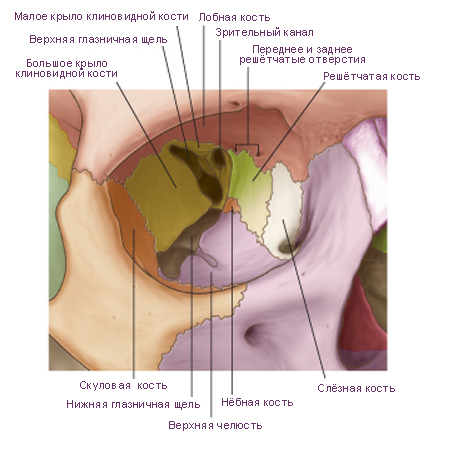
Seven bones contribute to the framework of each orbit. They are the maxilla, zygomatic, frontal, ethmoid, lacrimal, sphenoid, and palatine bones. Together they give the bony orbit the shape of a pyramid, with its wide base opening anteriorly onto the face, and its apex extending in a posteromedial direction. Completing the pyramid configuration are medial, lateral, superior, and inferior walls. |
 |
|
The apex of the pyramidal-shaped bony orbit is the optic foramen, while the base (the orbital rim) is formed:
• superiorly by the frontal bone;
• medially by the frontal process of the maxilla;
• inferiorly by the zygomatic process of the maxilla and the zygomatic bone;
• laterally by the zygomatic bone, the frontal process of the zygomatic bone, and the zygomatic process of the frontal bone.
|
|
Примечание:
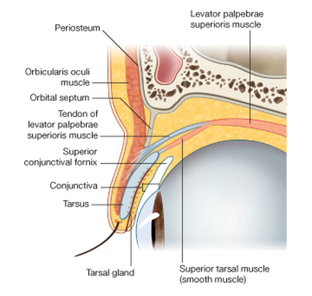
The upper and lower eyelids are anterior structures that, when closed, protect the surface of the eyeball.
The space between the eyelids, when they are open, is the palpebral fissure.
The layers of the eyelids, from anterior to posterior, consist of skin, subcutaneous tissue, voluntary muscle, the orbital septum, the tarsus, and conjunctiva.
The upper and lower eyelids are basically similar in structure except for the addition of two muscles in the upper eyelid. |
 |
|
|
Схема. A. Движения глазного яблока. B. Оси глазного яблока и глазницы = A. Movements of the eyeball. B. Axes of of the eyeball and orbit.
Модификация: Gray H., (1821–1865), Drake R., Vogl W., Mitchell A., Eds. Gray's Anatomy for Students. Churchill Livingstone, 2007, 1150 p.
см.: Анатомия человека: Литература. Иллюстрации |
 |
|
Примечание:
|
Of the seven muscles in the extrinsic group of muscles, one raises the eyelids, while the other six move the eyeball itself.
The movements of the eyeball, in three dimensions, are:
• elevation-moving the pupil superiorly;
• depression-moving the pupil inferiorly;
• abduction-moving the pupil laterally;
• adduction-moving the pupil medially;
• internal rotation-rotating the upper part of the pupil medially (or towards the nose);
• external rotation-rotating the upper part of the pupil laterally (or towards the temple).
Additionally, the axis of each orbit is directed slightly laterally from back to front, while each eyeball is directed anteriorly. Therefore, the pull of some muscles has multiple effects on the movement of the eyeball, while that of others has single effects. |
|
|
Схема. Артерии (A) и вены (B) глазницы и глазного яблока = A. Arterial supply to the orbit and eyeball. B. Venous drainage of the orbit and eyeball.
Модификация: Gray H., (1821–1865), Drake R., Vogl W., Mitchell A., Eds. Gray's Anatomy for Students. Churchill Livingstone, 2007, 1150 p.
см.: Анатомия человека: Литература. Иллюстрации |
 |
|
Примечание:
|
Arteries
The arterial supply to the structures in the orbit, including the eyeball, is by the ophthalmic artery. This vessel is a branch of the internal carotid artery, given off immediately after the internal carotid artery leaves the cavernous sinus. The ophthalmic artery passes into the orbit through the optic canal with the optic nerve.
In the orbit the ophthalmic artery initially lies inferior and lateral to the optic nerve. As it passes forward in the orbit it crosses superior to the optic nerve and proceeds anteriorly on the medial side of the orbit.
In the orbit the ophthalmic artery gives off numerous branches as follows:
• the lacrimal artery, which arises from the ophthalmic artery on the lateral side of the optic nerve, and passes anteriorly on the lateral side of the orbit, supplying the lacrimal gland, muscles, the anterior ciliary branch to the eyeball, and the lateral sides of the eyelid;
• the central retinal artery, which enters the optic nerve, proceeds down the center of the nerve to the retina, and is clearly seen when viewing the retina with an ophthalmoscope-occlusion of this vessel or of the parent artery leads to blindness;
• the long and short posterior ciliary arteries, which are branches that enter the eyeball posteriorly, piercing the sclera, and supplying structures inside the eyeball;
• the muscular arteries, which are branches supplying the intrinsic muscles of the eyeball;
• the supra-orbital artery, which usually arises from the ophthalmic artery immediately after it has crossed the optic nerve, proceeds anteriorly, and exits the orbit through the supra-orbital foramen with the supra-orbital nerve-it supplies the forehead and scalp as it passes across these areas to the vertex of the skull;
• the posterior ethmoidal artery, which exits the orbit through the posterior ethmoidal foramen to supply the ethmoidal air cells and nasal cavity;
• the anterior ethmoidal artery, which exits the orbit through the anterior ethmoidal foramen, enters the cranial cavity giving off the anterior meningeal branch, and continues into the nasal cavity supplying the septum and lateral wall, and ending as the dorsal nasal artery;
• the medial palpebral arteries, which are small branches supplying the medial area of the upper and lower eyelids;
• the dorsal nasal artery, which is one of the two terminal branches of the ophthalmic artery, leaves the orbit to supply the upper surface of the nose;
• the supratrochlear artery, which is the other terminal branch of the ophthalmic artery and leaves the orbit with the supratrochlear nerve, supplying the forehead as it passes across it in a superior direction.
Veins
There are two venous channels in the orbit, the superior and inferior ophthalmic veins.
The superior ophthalmic vein begins in the anterior area of the orbit as connecting veins from the supra-orbital vein and the angular vein join together. It passes across the superior part of the orbit, receiving tributaries from the companion veins to the branches of the ophthalmic artery and veins draining the posterior part of the eyeball. Posteriorly, it leaves the orbit through the superior orbital fissure and enters the cavernous sinus.
The inferior ophthalmic vein is smaller than the superior ophthalmic vein, begins anteriorly, and passes across the inferior part of the orbit. It receives various tributaries from muscles and the posterior part of the eyeball as it crosses the orbit.
The inferior ophthalmic vein leaves the orbit posteriorly by:
• joining with the superior ophthalmic vein;
• passing through the superior orbital fissure on its own to join the cavernous sinus; or
• passing through the inferior orbital fissure to join with the pterygoid plexus of veins in the infratemporal fossa. |
|
|
Примечание:
Innervation
Numerous nerves pass into the orbit and innervate structures within its bony walls. They include the optic nerve [II], the oculomotor nerve [III], the trochlear nerve [IV], the abducent nerve [VI], and autonomic nerves. Other nerves such as the ophthalmic nerve [V1] innervate orbital structures and then travel out of the orbit to innervate other regions. |
 |
|
Optic nerve
The optic nerve [II] is not a true cranial nerve, but rather an extension of the brain carrying afferent fibers from the retina of the eyeball to the visual centers of the brain. It is surrounded by the cranial meninges, including the subarachnoid space, which extend as far forward as the eyeball.
Any increase in intracranial pressure therefore results in increased pressure in the subarachnoid space surrounding the optic nerve. This may impede venous return along the retinal veins, causing edema of the optic disc (papilledema) which can be seen when the retina is examined using an ophthalmoscope.
The optic nerve leaves the orbit through the optic canal. It is accompanied in the optic canal by the ophthalmic artery.
Oculomotor nerve
The oculomotor nerve [III] leaves the anterior surface of the brainstem between the midbrain and the pons. It passes forward in the lateral wall of the cavernous sinus.
Just before entering the orbit the oculomotor nerve [III] divides into superior and inferior branches. These branches enter the orbit through the superior orbital fissure, lying within the common tendinous ring.
Inside the orbit the small superior branch passes upward on the lateral side of the optic nerve to innervate the superior rectus and levator palpebrae superioris muscles.
The large inferior branch divides into three branches:
• one passing below the optic nerve as it passes to the medial side of the orbit to innervate the medial rectus muscle;
• a second descending to innervate the inferior rectus muscle;
• the third descends as it runs forward along the floor of the orbit to innervate the inferior oblique muscle.
As the third branch descends, it gives off the branch to the ciliary ganglion. This is the parasympathetic root to the ciliary ganglion and carries preganglionic parasympathetic fibers that will synapse in the ciliary ganglion with postganglionic parasympathetic fibers. The postganglionic fibers are distributed to the eyeball through short ciliary nerves and innervate the sphincter pupillae and ciliary muscles.
Trochlear nerve
The trochlear nerve [IV] arises from the posterior surface of the midbrain, and passes around the midbrain to enter the edge of the tentorium cerebelli. It continues on an intradural path arriving in and passing through the lateral wall of the cavernous sinus just below the oculomotor nerve [III].
Just before entering the orbit, the trochlear nerve ascends, passing across the oculomotor nerve [III] and enters the orbit through the superior orbital fissure above the common tendinous ring. In the orbit the trochlear nerve [IV] ascends and turns medially, crossing above the levator palpebrae superioris muscle to enter the upper border of the superior oblique muscle.
Abducent nerve
The abducent nerve [VI] arises from the brainstem between the pons and medulla. It enters the dura covering the clivus and continues in a dural canal until it reaches the cavernous sinus.
The abducent nerve enters the cavernous sinus and runs through the sinus lateral to the internal carotid artery. It passes out of the sinus and enters the orbit through the superior orbital fissure within the common tendinous ring. Once in the orbit it passes out laterally to supply the lateral rectus muscle.
Postganglionic sympathetic fibers
Preganglionic sympathetic fibers arise from the upper segments of the thoracic spinal cord, mainly T1. They enter the sympathetic chain through white rami communicantes, and ascend to the superior cervical ganglion where they synapse with postganglionic sympathetic fibers.
The postganglionic fibers are distributed along the internal carotid artery and its branches.
The postganglionic sympathetic fibers destined for the orbit travel with the ophthalmic artery. Once in the orbit the fibers are distributed to the eyeball either by:
• passing through the ciliary ganglion, without synapsing, and joining the short ciliary nerves, which pass from the ganglion to the eyeball; or
• passing through long ciliary nerves to reach the eyeball.
In the eyeball postganglionic sympathetic fibers innervate the dilator pupillae muscle.
Ophthalmic nerve [V1]
The ophthalmic nerve [V1] is the smallest and most superior of the three divisions of the trigeminal nerve. This purely sensory nerve receives input from structures in the orbit and from additional branches on the face and scalp.
Leaving the trigeminal ganglion, the ophthalmic nerve [V1] passes forward in the lateral wall of the cavernous sinus inferior to the trochlear [IV] and oculomotor [III] nerves. Just before it enters the orbit it divides into three branches-the nasociliary, lacrimal, and frontal nerves. These branches enter the orbit through the superior orbital fissure with the frontal and lacrimal nerves outside the common tendinous ring, and the nasociliary nerve within the common tendinous ring.
Lacrimal nerve
The lacrimal nerve is the smallest of the three branches of the ophthalmic nerve [V1]. Once in the orbit it passes forward along the upper border of the lateral rectus muscle. It receives a branch from the zygomaticotemporal nerve, which carries parasympathetic and sympathetic postganglionic fibers for distribution to the lacrimal gland.
Reaching the anterolateral aspect of the orbit, the lacrimal nerve supplies the lacrimal gland, conjunctiva, and lateral part of the upper eyelid.
Frontal nerve
The frontal nerve is the largest branch of the ophthalmic nerve [V1] and receives sensory input from areas outside the orbit. Exiting the superior orbital fissure, this branch passes forward between the levator palpebrae superioris and the periorbita on the roof of the orbit. About midway across the orbit it divides into its two terminal branches-the supra-orbital and supratrochlear nerves:
• the supratrochlear nerve continues forward in an anteromedial direction, passing above the trochlea, exits the orbit medial to the supra-orbital foramen, and supplies the conjunctiva and skin of the upper eyelid and the skin on the lower medial part of the forehead;
• the supra-orbital nerve is the larger of the two branches, continues forward, passing between the levator palpebrae superioris muscle and the periorbita covering the roof of the orbit, exits the orbit through the supra-orbital notch and ascends across the forehead and scalp, supplying the upper eyelid and conjunctiva, the forehead, and as far posteriorly as the middle of the scalp.
Nasociliary nerve
The nasociliary nerve is intermediate in size between the frontal and lacrimal nerves and is usually the first branch from the ophthalmic nerve. It is most deeply placed in the orbit, entering the area within the common tendinous ring between the superior and inferior branches of the oculomotor nerve [III].
Once in the orbit, the nasociliary nerve crosses the superior surface of the optic nerve as it passes in a medial direction below the superior rectus muscle. Its first branch, the communicating branch with the ciliary ganglion (sensory root to the ciliary ganglion), is given off early in its path through the orbit.
The nasociliary nerve continues forward along the medial wall of the orbit, between the superior oblique and the medial rectus muscles, giving off several branches. These include:
• the long ciliary nerves, which are sensory to the eyeball but may also contain sympathetic fibers for pupillary dilation;
• the posterior ethmoidal nerve, which exits the orbit through the posterior ethmoidal foramen to supply posterior ethmoidal air cells and the sphenoidal sinus;
• the infratrochlear nerve, which distributes to the medial part of the upper and lower eyelids, the lacrimal sac, and skin of the upper half of the nose;
• the anterior ethmoidal nerve, which exits the orbit through the anterior ethmoidal foramen to supply the anterior cranial fossa, nasal cavity, and skin of the lower half of the nose.
Ciliary ganglion
The ciliary ganglion is a parasympathetic ganglion of the occulomotor nerve [III]. It is associated with the nasociliary branch of the ophthalmic nerve [V1] and is the site where preganglionic and postganglionic parasympathetic neurons synapse as fibers from this part of the autonomic division of the PNS make their way to the eyeball. The ciliary ganglion is also traversed by postganglionic sympathetic fibers and sensory fibers as they travel to the eyeball.
The ciliary ganglion is a very small ganglion, in the posterior part of the orbit immediately lateral to the optic nerve and between the optic nerve and the lateral rectus muscle. It is usually described as receiving at least two, and possibly three, branches or roots from other nerves in the orbit.
Parasympathetic root
As the inferior branch of the oculomotor nerve [III] passes the area of the ciliary ganglion, it sends a branch to the ganglion (the parasympathetic root). The parasympathetic branch carries preganglionic parasympathetic fibers, which enter the ganglion and synapse with postganglionic parasympathetic fibers within the ganglion.
The postganglionic parasympathetic fibers leave the ganglion through short ciliary nerves, which enter the posterior aspect of the eyeball around the optic nerve.
In the eyeball the parasympathetic fibers innervate:
the sphincter pupillae muscle, responsible for pupillary constriction;
the ciliary muscle, responsible for accommodation of the lens of the eye for near vision.
Sensory root
A second branch (the sensory root), passes from the nasociliary nerve to the ganglion. This branch enters the posterosuperior aspect of the ganglion, and carries sensory fibers, which pass through the ganglion and continue along the short ciliary nerves to the eyeball. These fibers are responsible for sensory innervation to all parts of the eyeball.
Sympathetic root
The third branch to the ciliary ganglion is the most variable. This branch, when present, is the sympathetic root and contains postganglionic sympathetic fibers from the superior cervical ganglion. These fibers travel up the internal carotid artery, leave the plexus surrounding the artery in the cavernous sinus, and enter the orbit through the common tendinous ring. In the orbit they enter the posterior aspect of the ciliary ganglion, cross the ganglion, and continue along the short ciliary nerves to the eyeball.
Sympathetic fibers to the eyeball may not enter the ganglion as a separate branch. The postganglionic sympathetic fibers may leave the plexus associated with the internal carotid artery in the cavernous sinus, join the ophthalmic nerve [V1] and distribute to the ciliary ganglion through the sensory root from the nasociliary nerve.
Whatever their path, postganglionic sympathetic fibers reach the eyeball and innervate the dilator pupillae muscle.
|
|
Схема. Глазное яблоко = Eyeball.
Модификация: Gray H., (1821–1865), Drake R., Vogl W., Mitchell A., Eds. Gray's Anatomy for Students. Churchill Livingstone, 2007, 1150 p.
см.: Анатомия человека: Литература. Иллюстрации |
 |
|
Примечание:
|
The globe-shaped eyeball occupies the anterior part of the orbit. Its rounded shape is disrupted anteriorly, where it bulges outward. This outward projection represents about one-sixth of the total area of the eyeball and is the transparent cornea. |
|
|
Примечание:
Ophthalmoscopy
Direct visualization of the postrenal (vitreous) chamber of the eye is possible in most clinical settings. It is achieved using an ophthalmoscope, which is a small battery-operated torch with a tiny lens that allows direct visualization of the postrenal (vitreous) chamber and the posterior wall of the eye through the pupil and the lens. It is sometimes necessary to place a drug directly onto the eye to dilate the pupil for better visualization.
The optic nerve is easily seen. The typical four branches of the retinal artery and the fovea are also seen. |
 |
|
Using ophthalmoscopy the physician can look for diseases of the optic nerve, vascular abnormalities, and changes within the retina
|
|
Примечание:
Ciliary body
Extending from the anterior border of the choroid is the ciliary body (Fig. 8.101). This triangular-shaped structure, between the choroid and the iris, forms a complete ring around the eyeball. Its components include the ciliary muscle and the ciliary processes (Fig. 8.103).
The ciliary muscle consists of smooth muscle fibers arranged longitudinally, circularly, and radially. Controlled by parasympathetics traveling to the orbit in the oculomotor nerve [III], these muscle fibers, on contraction, decrease the size of the ring formed by the ciliary body. The ciliary processes are longitudinal ridges projecting from the inner surface of the ciliary body. |
 |
|
Extending from them are zonular fibers attached to the lens of the eyeball, which suspend the lens in its proper position and collectively form the suspensory ligament of the lens.
Contraction of the ciliary muscle decreases the size of the ring formed by the ciliary body. This reduces tension on the suspensory ligament of the lens. The lens therefore becomes more rounded (relaxed) resulting in accommodation of the lens for near vision.
Ciliary processes also contribute to the formation of aqueous humor.
|
Таблица. Внутренние (собственные) мышцы глазного яблока = Intrinsic muscles of the eye.
c. 613.
Модификация: Gray H., (1821–1865), Drake R., Vogl W., Mitchell A., Eds. Gray's Anatomy for Students. Churchill Livingstone, 2007, 1150 p.
см.: Анатомия человека: Литература. Иллюстрации. |
№ |
Muscle |
Location |
Insertion |
Function |
| 1 |
Ciliary |
Muscle fibers in the ciliary body |
Parasympathetics from the oculomotor nerve [III] |
Constricts ciliary body, relaxes tension on lens, lens become more rounded |
| 2 |
Sphincter pupillae |
Circularly arranged fibers in the iris |
Parasympathetics from the oculomotor nerve [III] |
Constricts pupil |
| 3 |
Dilator pupillae |
Radially arranged fibers in the iris |
Sympathetics from the superior cervical ganglion (T1) |
Dilates pupil |
|
Литература. Иллюстрации. References. Illustrations
Щелкни здесь и получи доступ в библиотеку сайта! Click here and receive access to the reference library!
- Basbaum A.I., Shepherd G.M., Kaneko A., Westheimer J., Eds. The Senses: A Comprehensive Reference = Чувства. Справочник. 6-vol set, Academic Press, 2007, 4640 p. Иллюстрированное учебное пособие. .
Доступ к данному источнику = Access to the reference.
URL: http://www.tryphonov.ru/tryphonov/serv_r.htm#0 quotation
- Burnstock G., Sillito A.M., Eds., Nervous Control of the Eye = Нейрогенные механизмы управления в зрительной системе. Informa Health Care, 2000, 320 p. Иллюстрированное учебное пособие. Анатомия, гистология, физиология. .
Доступ к данному источнику = Access to the reference.
URL: http://www.tryphonov.ru/tryphonov/serv_r.htm#0 quotation
- Chalupa L.M., and Werner J.S., Eds. he Visual Neurosciences = Нейрофизиология зрения. Двухтомник. 2-vol set, The MIT Press, 2003, 1808 p. Сборник обзоров.
Доступ к данному источнику = Access to the reference.
URL: http://www.tryphonov.ru/tryphonov/serv_r.htm#0 quotation
- Dartt D.A., Ed. Encyclopedia of the Eye = Глаз. Энциклопедия. Четырёхтомник, 4 vol. set, Academic Press, 2010, 2344 p.
Иллюстрированное учебное пособие.
Доступ к данному источнику = Access to the reference.
URL: http://www.tryphonov.ru/tryphonov/serv_r.htm#0 quotation
- De Valois K.K. Seeing (Handbook of Perception and Cognition) = Зрение. Восприятие и познание. Academic Press, 2000, 392 p. Иллюстрированное учебное пособие. .
Доступ к данному источнику = Access to the reference.
URL: http://www.tryphonov.ru/tryphonov/serv_r.htm#0 quotation
- Faller A., Schuenke M., Eds. The Human Body = Тело человека. Thieme, 2004, 710 p.
Основные принципы анатомии и физиологии тела человека. Прекрасно иллюстрированный учебник для средних учебных заведений.
Доступ к данному источнику = Access to the reference. Атласы и учебники издательства Thieme.
URL: http://www.tryphonov.ru/tryphonov/serv_r.htm#0. quotation
- Friedman N.J., Kaiser P.K., Pineda R., Eds. The Massachusetts Eye and Ear Infirmary Illustrated Manual of Ophthalmology = Офтальмология, 3rd ed., Elsevier, 2011, ~86 MB.
Иллюстрированное учебное пособие. .
Доступ к данному источнику = Access to the reference.
URL: http://www.tryphonov.ru/tryphonov/serv_r.htm#0 quotation
- Gray H., (1821–1865), Drake R., Vogl W., Mitchell A., Eds. Gray's Anatomy for Students = Г. Грей: Анатомия для студентов. Churchill Livingstone, 2007, 1150 p.
Прекрасно иллюстрированное классическое учебное пособие и руководство, обновленное и дополненное коллективом современных авторов. В формате .chm. .
Доступ к данному источнику = Access to the reference.
URL: http://www.tryphonov.ru/tryphonov/serv_r.htm#0 quotation
- Gray H., (1821–1865), Standring S., Ed. Gray's Anatomy: The Anatomical Basis of Clinical Practice = Г. Грей: Анатомические основы клинической практики. 39th ed., Churchill Livingstone, 2008, 1600 p.
Прекрасно иллюстрированное классическое учебное пособие и руководство, обновленное и дополненное коллективом современных авторов. В формате .chm. .
Доступ к данному источнику = Access to the reference.
URL: http://www.tryphonov.ru/tryphonov/serv_r.htm#0 quotation
- Gray H., (1821–1865), Bannister L.H., Berry M.M., and Williams P.L., Eds. Gray's Anatomy: The Anatomical Basis of Medicine & Surgery = Г. Грей: Анатомические основы медицины и хирургии. 38th ed., Churchill Livingstone, 1995, 600 p.
Прекрасно иллюстрированное классическое учебное пособие и руководство, обновленное и дополненное коллективом современных авторов. В формате .pdf. .
Доступ к данному источнику = Access to the reference.
URL: http://www.tryphonov.ru/tryphonov/serv_r.htm#0 quotation
- Grierson I. Eye Book: Eyes and Eye Problems Explained = Глаз. Норма и патология. Liverpool University Press, 2000, 220 p. Иллюстрированное учебное пособие.
Доступ к данному источнику = Access to the reference.
URL: http://www.tryphonov.ru/tryphonov/serv_r.htm#0 quotation
- Grossman S., Ed. Porth's Pathophysiology = Патофизиология, Lippincott Williams & Wilkins, 2013, 1690 p.
Учебное пособие. .
Доступ к данному источнику = Access to the reference.
URL: http://www.tryphonov.ru/tryphonov/serv_r.htm#0 quotation
- Herranz R.M., Herran R.M.C., Eds. Ocular surface: anatomy and physiology, disorders, and therapeutic care = Поверхность глазного яблока. Анатомия, физиология, нарушения, лечение, Springer, 2005, 281 p.
Учебное пособие. .
Доступ к данному источнику = Access to the reference.
URL: http://www.tryphonov.ru/tryphonov/serv_r.htm#0 quotation
- Physiology of the Eye (CD-ROM) = Физиология глаза (диск), iKnow, 2005.
Интерактивный глаз.
Доступ к данному источнику = Access to the reference.
URL: http://www.tryphonov.ru/tryphonov/serv_r.htm#0 quotation
- Schlote T., Rohrbach J., Grueb M., Mielke J. Pocket atlas of ophthalmology = Офтальмология. Карманный атлас. Thieme, 2006, 214 p.
Учебное пособие. .
Доступ к данному источнику = Access to the reference.
URL: http://www.tryphonov.ru/tryphonov/serv_r.htm#0 quotation
- Valberg A. Light. Vision. Color = Свет. Зрение. Цвет. Wiley, 2005, 474 p. Иллюстрированное учебное пособие. .
Доступ к данному источнику = Access to the reference.
URL: http://www.tryphonov.ru/tryphonov/serv_r.htm#0 quotation
- Ward J.P.T., Linden R.W.A., Eds. Physiology at a Glance = Основы физиологии, 3rd ed., Wiley, 2013, 168 p.
Иллюстрированное учебное пособие..
Доступ к данному источнику = Access to the reference.
URL: http://www.tryphonov.ru/tryphonov/serv_r.htm#0 quotation
|
«Я У Ч Е Н Ы Й И Л И . . . Н Е Д О У Ч К А ?»
Т Е С Т В А Ш Е Г О И Н Т Е Л Л Е К Т А
Предпосылка:
Эффективность развития любой отрасли знаний определяется степенью соответствия методологии познания - познаваемой сущности.
Реальность:
Живые структуры от биохимического и субклеточного уровня, до целого организма являются вероятностными структурами. Функции вероятностных структур являются вероятностными функциями.
Необходимое условие:
Эффективное исследование вероятностных структур и функций должно основываться на вероятностной методологии (Трифонов Е.В., 1978,..., ..., 2015, …).
Критерий: Степень развития морфологии, физиологии, психологии человека и медицины, объём индивидуальных и социальных знаний в этих областях определяется степенью использования вероятностной методологии.
Актуальные знания: В соответствии с предпосылкой, реальностью, необходимым условием и критерием...
...
о ц е н и т е с а м о с т о я т е л ь н о:
— с т е п е н ь р а з в и т и я с о в р е м е н н о й н а у к и,
— о б ъ е м В а ш и х з н а н и й и
— В а ш и н т е л л е к т !
|
♥ Ошибка? Щелкни здесь и исправь ее! Поиск на сайте E-mail автора (author): tryphonov@yandex.ru
|